3-Step Eye Examination Simulator Kit - Professional Optometry Training Tool for Students & Doctors - Perfect for Clinical Practice & Medical Education
$825 $1500-45%
Free shipping on all orders over $50
7-15 days international
27 people viewing this product right now!
30-day free returns
Secure checkout
62480902
Guranteed safe checkout
DESCRIPTION
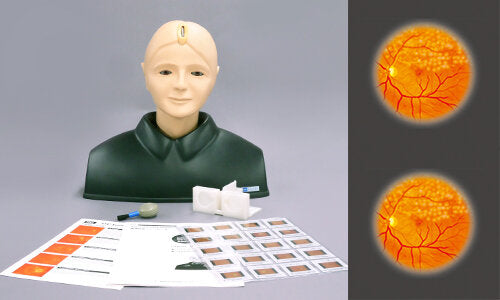
EYE Examination Simulator (3 Steps)
The EYE Examination Simulator is an innovative trainer for fundus examination, designed to allow examination of eyegrounds with the physician's own ophthalmoscope. Various cases can be set up for trainees using combinations of choice of slides, depth and pupil diameter. Soft and supple material allows hands-on simulation of real examination procedures, such as raising the eyelid.
Features
Lenses are used for a part of the eyeball, and reproduces the visual axis close to human.
It is possible to change the degree of dilation and contraction of the pupil in 3 steps (2, 3.5,5mm) offering different degrees of challenges. (*M82A is 2 steps; 3.5, 8mm)
The soft and supple material allows simulations of a real examination, in ways such as pulling up the eyelid.
Real clinical images
10 fundus slide cases of common eye diseases
You can examine the optic fundus with any ophthalmoscope available on the market. Provides you with the simulation of an actual examination.
Lens-Equipped eyeball units which simulates visual axis of human eye
Different pupil sizes can be set for challenge variation
Realistic raising of Eyelid Practice how to position your subdominant hand and your body against the patient.
Detectable Red Reflex
Most common pathologies with in 10 real clinical images
Adjustable fundus depth : Normal / Myopia / Hyperopia
Difference of KK-M82 (3 steps) and KK-M82A (2 steps)
Pupils of KK-M82A are designed larger than KK-M82. KK-M82A: 3.5mm / 8.0mm diameter. KK-M82: 2.0mm / 3.5mm / 5.0mm diameter.
This offers advantages for providing; - Dilated pupils when using mydriaticeye drops - Visual intelligibility for bigginers
Training skills / Applications
Funds examination with direct ophthalmoscope
Case / Pathology
1Normal fundus
2Hypertensive retinopathy:
Grade 3 arteriolar vasoconstriction
Grade 1 arteriolosclerosis
Hemorrhages and cotton wool spots
Simple vein concealment
3Diabetic retinopathy:
Microaneurysm, hemorrhages and hard exudates
4Papilloedema (chronic phase)
5Papilloedema (acute phase)
6Glaucomatous optic atrophy:
Glaucomatous optic disc cupping and nerve fiber defect